 |
|
|
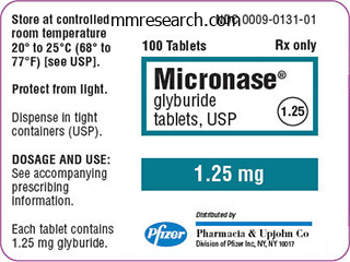
Micronase dosages: 5 mg, 2.5 mg
2.5 mg micronase generic free shippingThese tumors are extra typically infiltrative and regularly show foci of vascular and lymphatic invasion diabetes type 2 zwanger worden buy micronase 5 mg. Increasing tumor measurement uncomplicated diabetes definition micronase 2.5 mg buy visa, stage, high mitotic rate, diploma of mobile atypia, and necrosis seem to correlate with a poorer prognosis. Features of neuroendocrine differentiation could also be difficult to reveal in a few of these lesions, and never all tumors will give the anticipated positive reactions with immunohistochemical stains for neuroendocrine markers. The diagnosis is principally depending on routine gentle microscopy and recognition of the characteristic cytologic features. Subsequently, an additional variant of poorly differentiated neuroendocrine carcinoma was added to this roster: the large cell neuroendocrine carcinoma. Poorly differentiated neuroendocrine carcinomas of the lung account for about 20% of bronchogenic carcinomas and have a tendency to occur most frequently in sufferers between 50 and 70 years of age. Clinically, these tumors are extra usually situated centrally and present with symptoms of airway obstruction. The tumors commonly spread early into the mediastinum, giving rise to symptoms of superior vena cava syndrome, hoarseness, and dysphagia. Massive metastases to hilar and mediastinal lymph nodes are a common mode of presentation. Common extrathoracic websites of metastasis embrace bone, bone marrow, liver, and mind. Paraneoplastic syndromes similar to Cushing syndrome, the syndrome of inappropriate antidiuretic hormone secretion, and the EatonLambert syndrome are characteristically related to small cell lung cancer. Small Cell Carcinoma Small cell carcinoma is characterised by a proliferation of primitive-appearing, spherical to oval-shaped tumor cells that common two to thrice the dimensions of normal small lymphocytes. In the classical oat-cell variety, the tumor cells kind haphazardly arranged sheets of monotonous tumor cells which could be separated by thin fibrous septa and show in depth areas of necrosis. Nucleoli are usually inconspicuous or absent, and the tumor cells are characterized by brisk mitotic activity. In small endoscopic biopsies, the tumor cells will show a tendency to turn out to be markedly distorted and crushed, a discovering that traditionally has been related to small cell carcinoma but which will even be noticed in lymphoid neoplasms. A trabecular or ribbon-like arrangement of tumor cells, peripheral palisading of nuclei, and formation of rosette-like microacinar structures are rarely seen in small cell carcinoma and are options indicative of a better degree of differentiation. Mixed Small Cell�Large Cell Carcinoma Mixed small cell�large cell carcinomas are characterised by the presence of a subpopulation of enormous, undifferentiated tumor cells occurring singly or in small clusters inside an otherwise standard small cell carcinoma of the lung. A continuum of cell types is usually appreciated, ranging from typical small cells (oat cells) to the large cells. A massive cell element admixed with the small cells can be frequently noticed at metastatic websites of in any other case typical small cell carcinomas. Mixed small cell�large cell carcinoma appears to have a poorer survival and a more limited response to remedy than typical small cell carcinoma. B between areas of high-grade and intermediate-grade differentiation throughout the same tumor. In the "intermediate" subtype of small cell carcinoma, the cells appear larger, oval to polygonal, with extra marked nuclear pleomorphism, less stippling of chromatin, more prominent nucleoli, and more plentiful cytoplasm. Large cell neuroendocrine carcinoma has represented a controversial class for which the scientific implications, in addition to histopathologic standards for prognosis, have been solely more recently outlined. In general phrases, the overall histopathologic appearance of this lesion is that of a poorly differentiated non�small cell carcinoma. However, the mobile proliferation may be focally arranged in ribbons or cords admixed with rosettelike buildings, giving the lesion a vaguely neuroendocrine appearance. In addition, the tumor cells might show optimistic staining with neuroendocrine markers such as synaptophysin or chromogranin. This latter feature is much like that described in basaloid carcinoma of the lung, a variant of squamous cell carcinoma (see p 213). These tumors most likely originate from submucosal glands of the bronchi; however, not all of them occur in relationship with a bronchus. In uncommon circumstances, they could arise within the pulmonary parenchyma, in the periphery of the lung, with out direct connection to a bronchial construction. However, in latest times, several stories addressing their biologic conduct and spectrum of histopathologic features have been introduced. For instance, blended tumors, which symbolize the commonest tumor in salivary gland, are rarely seen in the lung. Other refined differences in the pathologic options and behavior of these lesions have additionally been famous. Clinically, they most often current as endobronchial lesions that trigger signs associated to bronchial obstruction, together with cough, dyspnea, and hemoptysis. Peripheral lesions usually have a tendency to be asymptomatic and therefore found by the way on routine chest radiograph. As a bunch, salivary gland�type tumors tend to behave as low-grade neoplasms with a good medical consequence when utterly resected. In particular, salivary gland� sort combined tumors, acinic cell carcinoma, low-grade mucoepidermoid carcinoma, and epithelial-myoepithelial carcinoma are curable by surgical excision alone. However, some exceptions exist; for example, adenoid cystic carcinomas may observe an aggressive course with distant unfold and high mortality, relying on the stage of the disease at the time of preliminary analysis. Tumors discovered to be at an advanced stage on the time of diagnosis will often prove fatal, impartial of the histologic options. Poorly differentiated salivary gland�type mixed tumors and mucoepidermoid carcinomas of high-grade histology may even present highly aggressive habits leading to demise due to widespread metastases. Adenoid Cystic Carcinoma Adenoid cystic carcinoma of the bronchus might reveal a selection of histologic growth patterns: (1) cribriform (cylindromatous), (2) tubular, and (3) stable. The islands of tumor cells are separated by fibrous bands and comprise cells with spherical nuclei displaying clear nuclear outlines and scant eosinophilic cytoplasm. The cystic areas are composed of two rows of cells, and mitotic figures are normally absent. The stable growth sample is probably probably the most uncommon and is characterised by related cells that kind diffuse sheets. Mitotic figures are found more regularly in association with the latter progress pattern. The only parameter that has been found to predict the prognosis for these tumors reliably is staging on the time of initial analysis. Immunohistochemical stains will identify both a glandular secretory and a myoepithelial component. Several morphologic development patterns have been identified in these tumors, together with acinar, cystic or papillocystic, nesting, and oncocytic. Some tumors may be composed predominantly of oncocytic cells displaying a strikingly nested development pattern, thus intently resembling a neuroendocrine neoplasm. Buy cheap micronase 2.5 mg onlineMoreover diabetes insipidus usg 2.5 mg micronase order with amex, it has been reported to present a variety of histologic subtypes diabetes test rite aid micronase 5 mg purchase visa, including typical, mobile, and melanocytic. The majority of lesions happen within the metaphysis of long bones (80%) and the vertebrae. The lesion is solely lytic, involves the metaphysis of an extended bone, is eccentric, and exhibits a blowout look with extension into gentle tissues. The soft tissue extension is normally restricted by a shell of periosteal new bone formation. The quantity of tissue acquired in the surgical pathology laboratory is often quite small compared with the size suggested radiographically. This is defined on the premise of the destruction of the spaces when the lesion is being removed. Microscopically, under low energy, aneurysmal bone cyst exhibits cysts of various sizes separated by septa. The septa are composed of loosely organized spindle cells with osteoclast-like large cells and capillary proliferation. Typically, just beneath the layer of cuboidal cells, a skinny layer of bone is formed that has been termed fiberosteoid. A, Vacuolated tumor cells show minimal atypia creating an look that resembles adipose tissue. Histologically, at low magnification, benign notochordal cell tumor fills the marrow spaces with out destroying the medullary or cortical bone. The tumor cells of benign notochordal cell tumor comprise small, spherical nuclei with minimal atypia surrounded by faintly eosinophilic or clear cytoplasm. Extensive vacuolation of the cytoplasm is a common feature, usually causing the lesion to be overlooked as benign fats. Benign notochordal cell tumors lack the myxoid matrix sometimes seen in chordoma, one of the most important options to distinguish these two tumors. Therefore keratin and brachyury are very helpful in separating it from adipose tissue, significantly because both lesions are immunoreactive with S-100 protein. So far, it appears that the biologic conduct of benign notochordal cell tumor is that of a benign lesion that must be handled conservatively or just noticed. Nevertheless, further studies are wanted to extra clearly perceive the biology of this lesion. A, Cystic areas surrounded by septa containing spindle cells without atypia and scattered multinucleate big cells. B, Fibrous septa composed of spindle cells and scattered multinucleate giant cells. Typically, new bone formation can additionally be current, creating an total appearance resembling myositis ossificans. The differential analysis primarily consists of giant cell tumor, simple cyst (see later discussion), and telangiectatic osteosarcoma. In a rare occasion, an abundance of giant cells could also be present, and the looks might recommend a large cell tumor. However, the lesional cells in aneurysmal bone cyst are slender and spindle shaped rather than round to oval as in a large cell tumor. Giant cell tumors happen in the ends of bones in grownup patients, whereas aneurysmal bone cysts occur within the metaphysis of youthful sufferers. When big cell tumor happens in the backbone, it entails the physique, whereas aneurysmal bone cyst involves the dorsal parts. However, at larger magnification, the tumor cells in telangiectatic osteosarcoma display apparent marked cytologic pleomorphism. Aneurysmal bone cyst is cytogenetically characterised by a recurrent rearrangement of chromosome band 17p13, more generally within the type of the balanced chromosomal translocation t(16;17)(q22;p13). Adjuvant therapies, including preoperative embolization, cryotherapy, and sclerotherapy, are generally used. Simple Bone Cyst Simple bone cysts happen in the proximal humerus and proximal femur in young kids. Boys are affected 1914 25 Tumors of the Osteoarticular System far more incessantly than girls, and the similar old presentation is that of a pathologic fracture. Radiography shows a purely lucent defect located centrally in the bone and increasing up to the epiphyseal plate. Grossly, the specimen shows a cystic lesion, with a thin fibrous lining, containing clear yellow fluid. Microscopically, skinny fibrous septa with an occasional osteoclast-type giant cell are seen. Amorphous eosinophilic material resembling cementum, however doubtless representing fibrin deposition, is usually seen within the cyst wall. The cyst wall of straightforward cyst is normally thinner and less mobile than aneurysmal bone cyst. The cementum-like materials is probably the most helpful histologic function that factors toward easy cyst. At instances, a lot histologic overlap is current that the final analysis depends on the radiographic features. The therapy of a simple bone cyst is aspiration of fluid and injection of methylprednisolone acetate. Surgical treatment is reserved for recurrent lesions after failure of injection with methylprednisolone acetate. Intraosseous Ganglion Intraosseous ganglia are nonneoplastic intraosseous lesions that are located at the ends of bones. Grossly and microscopically, they resemble the far more widespread ganglion cyst of sentimental tissues. Fewer than 5% of sufferers, predominantly those with polyostotic disease, present with skin pigmentation and endocrine abnormalities (McCune-Albright syndrome). The uncommon phenomenon of fibrous dysplasia associated with delicate tissue myxomas (Mazabraud syndrome) can also be more generally seen in polyostotic illness. The mutation normally results in a substitution of a histidine (R201H) or cysteine (R201C) for the arginine at place 201 of the protein. In the vast majority of instances, the findings on radiographs are those of a benign lesion with a differential diagnosis that features fibrous dysplasia. It can be a bit harder for radiologists to favor a benign diagnosis with confidence when fibrous dysplasia happens in rare places such as the spine. The basic radiographic description is that of a well-circumscribed lesion with a sclerotic rim and ground-glass look. Tumors situated within the long bones are often centered within the medulla and involve the metaphysis or diaphysis. Microscopically, fibrous dysplasia shows irregular trabeculae of woven bone inside fibrous tissue. Cytologically, the cells are often brief in contrast with the slender elongated cells characteristic of different fibrous lesions of bone.
5 mg micronase cheap otcNeuromuscular Hamartoma (Benign Triton Tumor) Neuromuscular hamartoma is an exceptionally rare lesion of infancy diabetes medications that cause hypoglycemia micronase 5 mg generic with mastercard,10 diabetes symptoms during exercise cheap 2.5 mg micronase with visa,eleven characterised by the congenital presence Solitary Circumscribed Neuroma (Palisaded Encapsulated Neuroma) Clinical Features Solitary circumscribed neuroma, first described in 1972 by Reed and colleagues,17 is sort of frequent and has usually been underrecognized by both clinicians and pathologists. It usually presents as a solitary painless skin nodule, less than 1 cm in diameter, within the "muzzle" or "butterfly" area of the face,17-19 though other mucocutaneous sites, including oral cavity20 and glans penis, may be affected. The peak incidence is between the ages of 30 and 60 years; no sex predilection is seen. No affiliation exists with any of the neurocristopathies, and the medical course is completely benign. The nodule often "pops" out of the encompassing tissue on the time of surgical procedure or gross examination. Note the dearth of a capsule superficially and the nerve of origin in the bottom right nook. The lesion itself is composed of bland eosinophilic spindle cells with poorly defined cell margins and small, wavy, hyperchromatic nuclei. The cells are arranged in fascicles set in a collagenous stroma and often separated by artifactual clefts. The relative circumscription of the lesion combined with the presence of a distinguished axonal component permits ready distinction from schwannoma or neurofibroma. Schwannoma (Neurilemmoma) Clinical Features Schwannoma is classically considered a benign, nonrecurring tumor of adulthood with no sex predilection,25,26 although distinctive instances (usually the mobile variant) may recur domestically. The anatomic distribution may be very broad, together with such diverse locations as the cranial nerves,27 bone,28 or the gastrointestinal tract, particularly the abdomen,29,30 but the overwhelming majority of instances develop in subcutaneous tissue, or less usually muscle, with a slight predilection for the distal extremities or head and neck area. With ever-increasing use of computed tomography and magnetic resonance imaging scans, small incidental schwannomas within the retroperitoneum are being identified more incessantly. At operation those tumors arising from an identifiable nerve can usually be separated simply therefrom, leaving no neurologic deficit. Most cases of solitary schwannoma are asymptomatic, and the majority are less than 5 cm in diameter. Convincingly recorded examples of benign schwannoma undergoing malignant change are very rare. Antoni A tissue is mobile and consists of monomorphic spindle-shaped Schwann cells, with poorly outlined eosinophilic cytoplasm and pointed basophilic nuclei, set in a variably collagenous stroma. Antoni B areas are also composed of Schwann cells, however their cytoplasm is inconspicuous, and the nuclei appear suspended in a copious myxoid, typically microcystic, matrix. A common feature, usually most distinguished in Antoni B areas, is the presence of blood vessels with thick hyaline partitions. Notably, schwannomas arising in the gastrointestinal or upper respiratory tracts tend distinctively to be unencapsulated, and those in the gastrointestinal tract have a distinguished peripheral lymphoid cuff. However, a fairly current examine demonstrated neurofilament-positive axons in nearly 50% of schwannomas, most frequently the standard and mobile subytpes. These modifications, that are initially focal, embody hyalinization, stromal hemorrhage, cystic change, and calcification. The point at which such tumors turn into classified as "ancient schwannoma" (see later discussion) is blurred. Immunohistochemically, schwannomas show diffuse and strong S-100 protein positivity. Bundles of long-spaced collagen (known as Luse bodies) are often seen within the stroma. By cytogenetic evaluation, most schwannomas present either monosomy 22 or loss of 22q material. First characterised in 1981, this variant of schwannoma has been mistaken for sarcoma (at least within the past) in as a lot as 30% of instances. A slight predominance in women is seen, and occasional instances have been related to neurofibromatosis. Although the tumor is normally encapsulated, some instances may be focally infiltrative and Variants of Benign Schwannoma Ancient Schwannoma. This very hyalinized, pseudovascular lesion contained only small foci of residual S-100�positive Schwann cells. It is necessary to notice that rare examples of mobile schwannoma could have a strikingly plexiform look. This unusual kind of schwannoma clinically tends to have an effect on barely younger patients than strange schwannoma. Each nodule is encapsulated, and mitoses could additionally be current, as in odd schwannoma. This uncommon variant of schwannoma usually arises in middle-aged adults and reveals a predilection for spinal nerve roots, although single instances at a extensive variety of net sites have been reported. Ultrastructurally, the tumor cells show typical Schwann cell options but additionally include melanosomes. Histologically, these lesions differ from ordinary schwannoma by being hypercellular virtually throughout (with an inconspicuous or only focal Antoni B component) and by showing a markedly fascicular. Mitoses may quantity as a lot as 10 per 10 hpf, and nuclear pleomorphism may be seen; the latter is degenerative (pyknotic) in nature and is often dissociated from the mitotic areas. Other common features are the presence of a thick fibrous capsule, during which there may be a dense lymphocytic infiltrate, and the discovering of fairly quite a few foamy (xanthomatous) cells within the tumor. Antoni A�type tissue (without palisading or hyaline vessels) and having a markedly whorled progress pattern. They seem not to be related to any neurocristopathy and show no tendency for local recurrence. Note the sometimes heavy pigmentation (A) and the quite vesicular, grooved nuclei of tumor cells (B). This is a uncommon morphologic variant of schwannoma, affecting primarily adults, which exhibits a predilection to arise in the gastrointestinal tract. However, extremely uncommon benign counterparts do appear to exist, although it seems possible that abnormalities). In some circumstances, distinction from metastatic melanoma may be very tough, if not inconceivable. In different cases, nevertheless, the histologic options of the primary tumor could seem deceptively benign. Although sometimes encapsulated, they lack the Antoni A or B zonation or hyaline vessels of typical schwannoma and include nests or trabeculae of eosinophilic epithelioid cells, often in a myxoid stroma. These lesions have primarily schwannian cytomorphology but have a whorled progress pattern and lack Antoni A-B zonation. This was the name formerly given to these lesions which might be nowadays acknowledged as intranodal myofibroblastoma (see Chapter 21). However, very uncommon, real circumstances of benign schwannoma arising in a lymph node do seem to exist. More hardly ever, solitary neurofibroma could occur in deep delicate tissue, usually in an axial location. In distinction to schwannoma, neurofibroma seems to originate within the endoneurium, and, in reality, a small subset of neurofibromas are entirely intraneural. Localized neurofibroma appears to show no tendency for native recurrence; actually, its combined cell content material (see later discussion) and apparent endoneurial origin recommend that it is probably not neoplastic but perhaps would be better considered hamartomatous. This problem is more likely to stay unresolved till a consensus definition of a neoplasm may be reached (see Chapter 1), because neurofibromas have been shown to be clonal,seventy five,76 regardless of morphologic evidence of their polytypic mobile populations.
Buy cheap micronase 5 mg on lineCoffin C M blood glucose 59 cheap micronase 5 mg on line, Rulon J diabetes 02190 micronase 2.5 mg generic fast delivery, Smith L, Bruggers C, White F V 1997 Pathologic options of rhabdomyosarcoma before and after therapy: a clinicopathologic and immunohistochemical analysis. Bergonse F N, Nico M M, Kavamura M I, Sotto M N 2002 Miliary osteoma of the face: a report of four circumstances and review of the literature. Fanburg-Smith J C, Bratthauer G L, Miettinen M 1999 Osteocalcin and osteonectin immunoreactivity in extraskeletal osteosarcoma: a examine of 28 instances. Parham D M 2001 Pathologic classification of rhabdomyosarcomas and correlations with molecular studies. Begin L R, Schurch W, LaCoste J, Hiscott J, Melnychuk D A 1994 Glycogen-rich clear cell rhabdomyosarcoma of the mediastinum. Furlong M A, Fanburg-Smith J C 2001 Pleomorphic rhabdomyosarcoma in kids: four cases within the pediatric age group. Folpe A L, McKenney J K, Bridge J A, Weiss S W 2002 Sclerosing rhabdomyosarcoma in adults: report of four circumstances of a hyalinizing, matrix-rich variant of rhabdomyosarcoma that could be confused with osteosarcoma, chondrosarcoma or angiosarcoma. Wang J, Tu X, Sheng W 2008 Sclerosing rhabdomyosarcoma: a clinicopathologic and immunohistochemical research of 5 cases. Seidal T, Kindblom L-G, Angervall L 1989 Rhabdomyosarcoma in middle-aged and elderly people. Anderson J, Gordon A, Pritchard-Jones K, Shipley J 1999 Genes, chromosomes and rhabdomyosarcoma. Rodriguez-Peralto J L, Lopez-Barea F, Sanchez-Herrara S, Atienza M 1994 Primary aneurysmal cyst of soppy tissues. Fetsch J F, Laskin W B, Miettinen M 2001 Superficial acral fibromyxoma: a clinicopathologic and immunohistochemical analysis of 37 instances of a particular soft tissue tumor with a predilection for the fingers and toes. Allen P W, Dymock R B, MacCormac W B 1988 Superficial angiomyxomas with and without epithelial elements. Fetsch J F, Laskin W B, Tavossoli F A 1997 Superficial angiomyxoma (cutaneous myxoma): a clinicopathologic examine of 17 instances arising within the genital region. Carney J A, Toorkey B C 1991 Myxoid fibroadenoma and allied situations (myxomatosis) of the breast. Steeper T A, Rosai J 1983 Aggressive angiomyxoma of the feminine pelvis and perineum. Iezzoni J C, Fechner R E, Wong L S, Rosai J 1995 Aggressive angiomyxoma in males: a report of 4 circumstances. Rosai J, Limas C, Husband E M 1984 Ectopic hamartomatous thymoma: a distinctive benign lesion of the lower neck. Marshall-Taylor C, Fanburg-Smith J C 2000 Hemosiderotic fibrohistiocytic lipomatous lesion: ten instances of a beforehand undescribed fatty lesion of the foot/ankle. Folpe A L, Weiss S W 2004 Pleomorphic hyalinizing angiectatic tumor: evaluation of forty one circumstances supporting evolution from a distinctive precursor lesion. Enzinger F M, Weiss S W, Liang C Y 1989 Ossifying fibromyxoid tumor of soft components. Miettinen M, Finnell V, Fetsch J F 2008 Ossifying fibromyxoid tumor of sentimental parts-a clinicopathologic and immunohistochemical examine of 104 circumstances with long-term follow-up and a crucial evaluation of the literature. Folpe A L, Weiss S W 2003 Ossifying fibromyxoid tumor of sentimental parts: a clinicopathologic study of 70 circumstances with emphasis on atypical and malignant variants. Fisher C, Miettinen M 1997 Parachordoma: a clinicopathologic and immunohistochemical research of 4 cases of an uncommon soft tissue neoplasm. Folpe A L, Agoff S N, Willis J, Weiss S W 1999 Parachordoma is immunohistochemically and cytogenetically distinct from axial chordoma and extraskeletal myxoid chondrosarcoma. Smith B C, Ellis G L, Meis-Kindblom J M, Williams S B 1995 Ectomesenchymal chondromyxoid tumor of the anterior tongue. A distinct fibrohistiocytic tumor of kids and younger adults simulating a vascular neoplasm. Fanburg-Smith J C, Miettinen M 1999 Angiomatoid "malignant" fibrous histiocytoma: a clinicopathologic study of 158 instances and additional exploration of the myoid phenotype. Roth J A, Enzinger F M, Tannenbaum M 1975 Synovial sarcoma of the neck: a follow-up examine of 24 instances. Al-Daraji W, Lasota J, Foss R, Miettinen M 2009 Synovial sarcoma involving the top: analysis of 36 circumstances with predilection to the parotid and temporal regions. Folpe A L, Schmidt R A, Chapman D, Gown A M 1998 Poorly differentiated synovial sarcoma: immunohistochemical distinction from primitive neuroectodermal tumors and excessive grade malignant peripheral nerve sheath tumors. Smith T A, Machen S K, Fisher C, Goldblum J R 1999 Usefulness of cytokeratin subsets for distinguishing monophasic synovial sarcoma from malignant peripheral nerve sheath tumor. Sandberg A A, Bridge J A 2002 Updates on the cytogenetics and molecular genetics of bone and gentle tissue tumors. Bleeker J S, Quevedo J F, Folpe A L 2012 "Malignant" perivascular epithelioid cell neoplasm: risk stratification and treatment strategies. Halling A C, Wollan P C, Pritchard D J, Vlasak R, Nascimento A G 1996 Epithelioid sarcoma: a clinicopathologic review of 55 circumstances. Flucke U, Hulsebos T J, van Krieken J H, Mentzel T 2010 Myxoid epithelioid sarcoma: a diagnostic challenge. Miettinen M, Fanburg-Smith J C, Virolainen M, Shmookler B M, Fetsch J F 1999 Epithelioid sarcoma: an immunohistochemical analysis of 112 classical and variant instances and a dialogue of the differential prognosis. Foschini M P, Eusebi V 1994 Alveolar soft-part sarcoma: a brand new kind of rhabdomyosarcoma Christopherson W M, Foote F W, Stewart F W 1952 Alveolar soft-part sarcomas: structurally characteristic tumors of unsure histogenesis. Flieder D B, Moran C A, Suster S 1997 Primary alveolar soft-part sarcoma of the mediastinum: a clinicopathological and immunohistochemical examine of two cases. Wang N P, Bacchi C E, Jiang J J, McNutt M A, Gown A M 1996 Does alveolar soft-part sarcoma exhibit skeletal muscle differentiation An immunocytochemical and biochemical study of myogenic regulatory protein expression. Meis-Kindblom J M, Bergh P, Gunterberg B, Kindblom L G 1999 Extraskeletal myxoid chondrosarcoma: a reappraisal of its morphologic spectrum and prognostic components based mostly on 117 cases. Massi D, Beltrami G, Capanna R, Franchi A 2004 Histopathological reclassification of extremity pleomorphic delicate tissue sarcoma has clinical relevance. DeBlois G, Wang S, Kay S 1986 Microtubular aggregates inside tough endoplasmic reticulum: an uncommon ultrastructural function of extraskeletal myxoid chondrosarcoma. Parham D M, Weeks D A, Beckwith J B 1994 the clinicopathologic spectrum of putative extrarenal rhabdoid tumors. An analysis of forty two cases studied with immunohistochemistry and/or electron microscopy. Fanburg-Smith J C, Hengge M, Hengge U R, Smith S, Miettinen M 1998 Extrarenal rhabdoid tumors of sentimental tissue: a clinicopathologic and immunohistochemical examine of 18 instances. Madigan C E, Armenian S H, Malogolowkin M H, Mascarenhas L 2007 Extracranial malignant rhabdoid tumors in childhood: the Childrens Hospital Los Angeles experience. An estimated 2890 new circumstances of primary bone sarcoma happen in the United States per 12 months in contrast with 226,160 new circumstances of lung most cancers and 229,060 new circumstances of breast most cancers. This understandably results in uncertainty in diagnosing even moderately simple circumstances. One of crucial things for pathologists to bear in mind is that a staff method is essential in the management of a affected person with a bone tumor. In 1958, Jaffe2 identified the significance of cooperation among the surgeon, the radiologist, and the pathologist in diagnosing a lesion of bone.
Micronase 2.5 mg buy low costDespite the invention of numerous molecular and cytogenetic markers diabetes diet how many carbs per day discount 2.5 mg micronase fast delivery, cell kind stays an impartial prognostic factor in most research blood sugar test strips 2.5 mg micronase. Spindle A cells are elongated and include a nucleus with a central fold (as seen in Brenner tumors of the ovary). The nuclei of spindle B cells lack a central fold and feature a distinguished nucleolus. Epithelioid cells sometimes have abundant cytoplasm, open nuclei, and large, pleomorphic nucleoli. Proliferation In many research, the number of mitotic figures recognized in forty high-power (40�) fields (hpf) is recorded as a prognostic attribute. Proliferation indices utilizing Ki67 even have prognostic significance and may be recorded rather than mitotic counts. Tumor-infiltrating lymphocytes Tumors showing >100 lymphocytes per 20 (40�) hpf have been proven to carry a poorer prognosis than these with <100 per 20 hpf. Vasculogenic mimicry patterns this tumor characteristic is well detectable and extremely reproducible between pathologists. Cytogenetics Monosomy three and different cytogenetic abnormalities have been related to opposed consequence. This approach has been proven to discriminate between patients with an excellent consequence and those at high danger of metastasis. Cytogenetics Multiple cytogenetic abnormalities, most notably monosomy three, have been associated with an opposed end result in uveal melanoma. The loops have been proven to be constructive for laminin and heparin sulfate proteoglycan. They have been shown to conduct plasma and probably pink blood cells and are formed by extremely invasive tumor cells via a course of generally known as vasculogenic mimicry. Early data point out that tumor cells with this profile are distributed homogeneously all through the tumor, not like the heterogeneous distribution of tumor cells with monosomy 3. The statement that no patients with a category 1 gene expression profile die of metastatic melanoma raises the query of whether this assay has recognized a molecular profile of a uveal melanocytic nevoid lesion99 (see the introduction to this chapter). Folberg R, McLean I W, Zimmerman L E 1984 Conjunctival acquired melanosis and malignant melanoma. Folberg R, McLean I W, Zimmerman L E 1985 Primary acquired melanosis of the conjunctiva. The cells are polygonal, the nuclei are pleomorphic, and the nucleoli are prominent. Sinard J H 1999 Immunohistochemical distinction of ocular sebaceous carcinoma from basal cell and squamous cell carcinoma. Tahery D P, Goldberg R, Moy R L 1992 Malignant melanoma of the eyelid: a report of eight cases and a review of the literature. Scott I U, Karp C L, Nuovo G J 2002 Human papillomavirus 16 and 18 expression in conjunctival intraepithelial neoplasia. Rao N A, Font R L 1976 Mucoepidermoid carcinoma of the conjunctiva: a clinicopathologic examine of five cases. Huntington A C, Langloss J M, Hidayat A A 1990 Spindle cell carcinoma of the conjunctiva: an immunohistochemical and ultrastructural examine of six instances. Jakobiec F A, Bhat P, Colby K A 2010 Immunohistochemical studies of conjunctival nevi and melanomas. Ackerman A B, Sood R, Koenig M 1991 Primary acquired melanosis of the conjunctiva is melanoma in situ. Damato B, Coupland S E 2008 Conjunctival melanoma and melanosis: a reappraisal of terminology, classification and staging. Clin Experiment Ophthalmol 36: 786-795 29 Tumors of the Eye and Ocular Adnexa 2115 38. Griffith W R, Green W R, Weinstein G W 1971 Conjunctival malignant melanoma originating in acquired melanosis sine pigmento. Folberg R, McLean I W 1986 Primary acquired melanosis and melanoma of the conjunctiva: terminology, classification and biologic behavior. Jakobiec F A, Folberg R, Iwamoto T 1989 Clinicopathologic traits of premalignant and malignant melanocytic lesions of the conjunctiva. Mehta M, Jakobiec F, Fay A 2009 Idiopathic fibroinflammatory illness of the face, eyelids, and periorbital membrane with immunoglobulin G4-positive plasma cells. Kivela T 1999 Trilateral retinoblastoma: a meta-analysis of hereditary retinoblastoma associated with main ectopic intracranial retinoblastoma. Juarez C P, Tso M O 1980 An ultrastructural research of melanocytomas (magnocellular nevi) of the optic disk and uvea. Folberg R, Hendrix M J, Maniotis A J 2000 Vasculogenic mimicry and tumor angiogenesis. Folberg R 2010 the molecular classification of uveal melanocytic lesions: an necessary discovery in context. Robertson D M, Winkelmann R K 1984 Ophthalmic features of necrobiotic xanthogranuloma with paraproteinemia. Levin L A, Jakobiec F A 1992 Optic nerve tumors of childhood: a decision-analytical approach to their diagnosis. Stern J, Jakobiec F A, Housepian E M 1980 the architecture of optic nerve gliomas with and with out neurofibromatosis. Broughton W L, Zimmerman L E 1978 A clinicopathologic study of 56 cases of intraocular medulloepitheliomas. Bunt A H, Tso M O 1981 Feulgen-positive deposits in retinoblastoma: incidence, composition, and ultrastructure. Kopelman J E, McLean I W, Rosenberg S H 1987 Multivariate analysis of danger components for metastasis in retinoblastoma treated by enucleation. The inner ear consists of a particular form of inert bone, a virtually nonmitotic sensory space, and nerves. Its tumors are mainly these of Schwann cell lineage, the one comparatively labile cells in that area. The center ear is lined by a simple flat epithelium, however its capability to respond briskly to irritation by the production of glands (mucosal metaplasia) explains why adenoma is the commonest center ear neoplasm. The exterior ear is a specialised appendage of the skin, and its neoplasms largely replicate the vary seen in other cutaneous areas. Classification of those embryologic anomalies was first launched in a classic work by Walter Payton Work, M. The fluid content material is cloudy to purulent and viscid and often contains necrotic particles. In many cases, serial or deeper sections could also be required to identify the cartilage or adnexal tissue. Work kind I lesions are of ectodermal origin only and present a unilocular cyst lined by stratified squamous epithelium or ciliated respiratory epithelium; they tend to be preauricular and parallel to the Ancillary Tests In general, fine-needle aspiration is really helpful in the evaluation of all neck cysts. An epidermal inclusion cyst is histologically identical, thus requiring clinical information about a sinus or fistula, or the presence of mesodermal elements (adnexal buildings and/or cartilage) to make the excellence. A benign lymphoepithelial cyst exhibits intimate blending of epithelium with lymphoid tissue and is usually within or adjacent to the parotid gland. Folliculitis tends to be extra superficial, centered across the hair shaft or follicle (root) and lacks a sinus or fistula. Micronase 2.5 mg purchase on lineGrossly diabetes in dogs expense buy micronase 5 mg, gangliogliomas are relatively well demarcated and characteristically exhibit a cyst-mural nodule architecture diabetes new medications 2010 micronase 2.5 mg with mastercard. Histologically, gangliogliomas are composed of a mix of neoplastic ganglion cells and glial cells. The ganglion cells are dysmorphic, are irregular in shape, and may present considerable variation in size and form. Bizarre and binucleate types are current, however the latter may be uncommon in some tumors. Smears of ganglioglioma readily reveal mixed astrocytic and ganglionic populations in a fibrillary stroma. Eosinophilic granular or hyaline our bodies are commonly intermixed throughout the glial element. In tissue sections, the mixed population of neoplastic astrocytes and irregular ganglion cells is often embedded in a fibrovascular stroma. In this temporal lobe ganglioglioma, the astrocytic part resembles a pilocytic astrocytoma. Bielschowsky silver impregnation readily demonstrates the abnormally oriented and shaped leashes of neuritic processes from the ganglion cells. The stromal element is variable and ranges from delicate fibrovascular components admixed with an plentiful glial population to a relatively intense desmoplastic reaction that just about obscures the glial matrix. On occasion, glomeruloid capillaries and/or hyalinized vessels are sufficiently plentiful as to mimic a vascular malformation. Ki67 labeling index is low in the great majority of gangliogliomas, and it seems to be confined to the glial part. A, the glial component of this anaplastic ganglioglioma is composed of small cells with scant glial cytoplasm and hyperchromatic nuclei. B, In the identical tumor, ganglion cells were readily identified by silver impregnation stain. Clinically, anaplastic transformation ought to be suspected in sufferers with a latest exacerbation of continual seizure activity and increasing T2 signal depth and distinction enhancement with neuroimaging. Desmoplastic Infantile Ganglioglioma and Desmoplastic Cerebral Astrocytoma of Infancy, World Health Organization Grade I Clinical Features. The tumors sometimes current as a very large supratentorial tumor, with strong and cystic parts. The majority of tumors manifest medical signs between 2 and 24 months of age (mean 6 months; median 4 months) with a slight male predominance. The frontoparietal regions are most commonly involved, however the tumors may also come up in different hemispheric sites; mixed sites are fairly common. Confinement to deep buildings or communication with the ventricular system is rare and has been described in solely very occasional circumstances. Reticulin deposition is a notable function of these neoplasms, which corresponds, partially, to the manufacturing of basal lamina by the neoplastic astrocytes. The density of the reticulin varies from diffuse accumulations (A) to compact islands (B) scattered among extra densely cellular blended, differentiated populations (C) or primitive neural cells (D). Immunohistochemistry for glial and neuronal cytoskeletal proteins demonstrates the divergent differentiation of the heterogeneous neuroepithelial cells. Tumors with similar clinical and radiologic options, however restricted to astrocytic differentiation, have been termed desmoplastic cerebral astrocytomas of infancy. The strong portion is either isodense or barely hyperdense with marked enhancement after administration of vascular distinction media, especially in areas of apposition to the meningeal surface. On T2-weighted images, the cysts sometimes have elevated signal depth whereas the solid element is often heterogeneous with variable intensities. The hallmark histopathologic function of those tumors is plentiful, typically dense desmoplasia, which confers a attribute firmness to these neoplasms. This may be particularly prominent at the leptomeningeal interface and is associated with collagen deposition. Microscopically, the desmoplasia is marked by deposition of a dense stroma in combination with neuroepithelial and fibroblastic elements. Special stains can show wavy, dense deposition of reticulin-positive stroma with collagen fibrils investing both cellular elements. The distribution of the astrocytic and neuronal elements in every tumor is often heterogeneous and correlates, partially, with the density of the stroma. Overall, the astrocytic cells comprise the most conspicuous neural element, particularly in regions with the most ample desmoplasia. The neuronal parts are extremely heterogeneous, starting from atypical ganglion-like types to the more frequent smaller polygonal cells. Mitoses and micronecrosis, if present, are most commonly current in affiliation with the primitive cell populations. The nodules are composed of a combination of "oligodendrocyte-like cells," astrocytes, and neuronal cells embedded in a mucinous and microcystic matrix. Between the nodules is a typical "specific element" whereby neurons lie embedded in a mucoid matrix. The neuronal element is generally composed of huge cells floating within the mucinous matrix. Two examples of typical dysembryoplastic neuroepithelial tumor at low magnification show a mildly mobile neoplasm with microcystic modifications throughout the superficial cortex (A and B). Considering the numerous morphology of those lesions, sufficient sampling of intact specimens is diagnostically essential. Nonrepresentative or minute specimens often immediate misguided diagnoses of oligodendroglioma or oligoastrocytomas. Tumor recurrence has been noticed in cases with incomplete initial surgical resection, and malignant transformation has been reported in only some cases. Papillary Glioneuronal Tumor, World Health Organization Grade I Clinical Features. Papillary glioneuronal tumors are uncommon glioneuronal tumors that happen over a broad age range and show no sex predilection. Papillary glioneuronal tumors are characterised by a pseudopapillary structure consisting of hyalinized vessels surrounded by a single layer of pseudostratified, small, cuboidal glial cells. In the intervening regions between the papillary constructions are focal collections or sheets of neuronal cells starting from neurocytes to small ganglion cells. The blended cellular elements are composed of oligodendrocytelike cells, astrocytes, and neurons in variable proportions; nonetheless, the oligodendrocyte-like component is most often predominant. Papillary glioneuronal tumors sometimes present neurocytic-like cells arranged around vessels forming pseudopapillary structures. The mixed glioneuronal differentiation is highlighted by neuronal related markers, particularly synaptophysin in the perivascular areas and interpapillary areas. Neurocytes and ganglion-like cells are highlighted by NeuN and neurofilament expression. Although a small number of instances have been reported to date, follow-up data point out that papillary glioneuronal tumors are benign in behavior. Rosette-forming tumor of the fourth ventricle is a uncommon glioneuronal tumor characteristically located within the midline occupying the fourth ventricle and involving the cerebellar vermis. The tumors may arise at any age (mean age at presentation 33 years; vary 12-59 years)407 with no significant intercourse predilection. Cheap micronase 2.5 mg amexThe difficulties stem from the morphologic overlap among the many different tumor sorts and difficulties in proper evaluation of the tumor borders in biopsies blood sugar after meals order micronase 2.5 mg. In the absence of extra definitive diagnostic features diabetes prevention best practices micronase 5 mg purchase with amex, only a descriptive diagnosis of "salivary gland neoplasm" may be made, and complete excision or a larger biopsy sample is required to arrive at a definitive prognosis. Frommer J 1977 the human accent parotid gland: its incidence, nature, and significance. Cleary K R, Batsakis J G 1990 Lymphoepithelial cysts of the parotid area: a new face on an old lesion. Krech R, Zerban H, Bannasch P 1981 Mitochondrial anomalies in renal oncocytes induced in rat by N-nitrosomorpholine. Hickman R E, Cawson R A, Duffy S W 1984 the prognosis of specific forms of salivary gland tumors. Laskawi R, Schott T, Schroder M 1998 Recurrent pleomorphic adenomas of the parotid gland: medical analysis and long-term follow-up. McGregor A D, Burgoyne M, Tan K C 1988 Recurrent pleomorphic salivary adenoma-the relevance of age at first presentation. Auclair P L, Ellis G L 1996 Atypical options in salivary gland mixed tumors: their relationship to malignant transformation. Coleman H, Altini M 1999 Intravascular tumour in intra-oral pleomorphic adenomas: a diagnostic and therapeutic dilemma. Pagni F, Fuertes Zarate A, Ren S 2010 Neural invasion in pleomorphic adenoma of the salivary gland. Lam P W, Chan J K, Sin V C 1997 Nasal pleomorphic adenoma with skeletal muscle differentiation: potential misdiagnosis as rhabdomyosarcoma. Takeda Y, Satoh M, Nakamura S 2004 Pigmented pleomorphic adenoma, a novel melanin-pigmented benign salivary gland tumor. Lee K C, Chan J K, Chong Y W 1992 Ossifying pleomorphic adenoma of the maxillary antrum. Am J Surg Pathol 24: 1159-1164 Manucha V, Ioffe O B 2008 Metastasizing pleomorphic adenoma of the salivary gland. Clin Orthop Relat Res 308: 192-198 Sampson B A, Jarcho J A, Winters G L 1998 Metastasizing mixed tumor of the parotid gland: a rare tumor with unusually speedy development in a cardiac transplant patient. Eur J Surg Oncol 24: 452-455 Eneroth C M, Zetterberg A 1974 Malignancy in pleomorphic adenoma. Head Neck Pathol 6: 1-9 Lewis J E, Olsen K D, Sebo T J 2001 Carcinoma ex pleomorphic adenoma: pathologic analysis of seventy three circumstances. Histopathology forty six: 144-152 Ellis G L, Auclair P L 1996 Tumors of the salivary glands. Oral Oncol 38: 107-110 Tortoledo M E, Luna M A, Batsakis J G 1984 Carcinomas ex pleomorphic adenoma and malignant combined tumors. Oral Surg Oral Med Oral Pathol Oral Radiol Endod 97: 613-619 Weber A, Langhanki L, Schutz A et al. J Oral Pathol Med forty: 460-466 Seifert G, Sobin L H 1991 Histological typing of salivary gland tumours. World Health Organization international histological classification of tumours, 2nd ed. Springer-Verlag, New York Muller S, Barnes L 1996 Basal cell adenocarcinoma of the salivary glands. Cancer seventy eight: 2471-2477 Fonseca I, Soares J 1996 Basal cell adenocarcinoma of minor salivary and seromucous glands of the pinnacle and neck area. J Laryngol Otol 103: 756-759 Ellis G L, Wiscovitch J G 1990 Basal cell adenocarcinomas of the major salivary glands. J Laryngol Otol 107: 862-864 Ellis G L, Auclair P L 1991 Basal cell adenocarcinoma. Saunders, Philadelphia, p 585-661 Murty G, Welch A, Soames J V 1990 Basal cell adenocarcinoma of the parotid gland. J Laryngol Otol 104: 150-151 Williams S B, Ellis G L, Auclair P L 1993 Immunohistochemical evaluation of basal cell adenocarcinoma. Oral Surg Oral Med Oral Pathol Oral Radiol Endod 87: 485-492 Dardick I, Thomas M J, van Nostrand A W 1989 Myoepithelioma- new ideas of histology and classification: a lightweight and electron microscopic examine. Virchows Arch A Pathol Anat Histopathol 416: 25-42 Dardick I 1995 Myoepithelioma: definitions and diagnostic criteria. American Registry of Pathology/Armed Forces Institute of Pathology, Washington, D. Histopathology 27: 1-9 Hornick J L, Fletcher C D 2003 Myoepithelial tumors of soft tissue: a clinicopathologic and immunohistochemical study of 101 circumstances with analysis of prognostic parameters. Hum Pathol 23: 748-754 Franquemont D W, Mills S E 1993 Plasmacytoid monomorphic adenoma of salivary glands. Oral Surg Oral Med Oral Pathol Oral Radiol Endod eighty one: 655-664 Hashimoto K, Yamamoto H, Shiratsuchi H et al. Cancer seventy nine: 2314-2319 Deguchi H, Hamano H, Hayashi Y 1993 c-myc, ras p21 and p53 expression in pleomorphic adenoma and its malignant type of the human salivary glands. Histopathology 51: 362-371 Altini M, Coleman H, Kienle F 1997 Intra-vascular tumour in pleomorphic adenomas-a report of four instances. J Laryngol Otol 104: 360-361 Kwon M Y, Gu M 2001 True malignant mixed tumor (carcinosarcoma) of parotid gland with uncommon mesenchymal component. Arch Pathol Lab Med a hundred twenty five: 812-815 Gnepp D R 1993 Malignant blended tumors of the salivary glands: a evaluate. Pathol Annu 28(Pt 1): 279-328 Batsakis J G, Luna M A, el-Naggar A K 1991 Basaloid monomorphic adenomas. Histopathology sixty one: 921-929 Dardick I, Daley T D, van Nostrand A W 1986 Basal cell adenoma with myoepithelial cell-derived "stroma": a new main salivary gland tumor entity. Am J Surg Pathol 26: 778-783 Edwards P C, Bhuiya T, Kelsch R D 2004 Assessment of p63 expression in the salivary gland neoplasms adenoid cystic 111. Savera A T, Gown A M, Zarbo R J 1997 Immunolocalization of three novel smooth muscle-specific proteins in salivary gland pleomorphic adenoma: assessment of the morphogenetic position of myoepithelium. Di Palma S, Guzzo M 1993 Malignant myoepithelioma of salivary glands: clinicopathological options of ten circumstances. Kane S V, Bagwan I N 2010 Myoepithelial carcinoma of the salivary glands: a clinicopathologic examine of fifty one cases in a tertiary cancer center. Shintaku M, Honda T 1997 Identification of oncocytic lesions of salivary glands by anti-mitochondrial immunohistochemistry. Gilcrease M Z, Nelson F S, Guzman-Paz M 1998 Tyrosine-rich crystals related to oncocytic salivary gland neoplasms. Ellis G L 1998 Clear cell neoplasms in salivary glands: clearly a diagnostic challenge. Maiorano E, Altini M, Favia G 1997 Clear cell tumors of the salivary glands, jaws, and oral mucosa. McMillan M D, Smith C J, Smillie A C 1993 Canalicular adenoma: report of five instances with ultrastructural observations.
Micronase 2.5 mg cheap on-lineS-100 protein is variably demonstrated in 20% to 50% jurnal diabetes mellitus type 2 micronase 5 mg, notably within the fibroblastic variant diabetes symptoms cats micronase 5 mg cheap with mastercard. The incidence of cytokeratin(s) immunoreactivity is approximately 20% throughout the whole group of meningiomas, however it might method 40% within the epithelial variants. Claudin-1, a good junction�associated protein, has been also demonstrated in about 50% of meningiomas and may be useful in distinguishing meningiomas from histologic mimics. Meningiomas are graded on the basis of histologic anaplasia and potential for aggressive habits (Table 26-9). In addition, special variants of meningiomas are acknowledged due to their specific potential for aggressive habits as highlighted beforehand. In addition, warning must be taken in grading meningiomas with a historical past of presurgical tumor embolization, a process performed in lots of the cases. Embolization induces tumoral necrosis with related reparative nuclear atypia, increased nucleolar measurement, and enhanced proliferation. Benign tumors on this class embrace chondroma, osteochondroma, osteoma, and lipoma. The most regularly encountered sarcomas include the so-called hemangiopericytoma, chondrosarcoma, and rhabdomyosarcoma. It is especially relevant to notice that hemangiopericytoma is considered a nonmeningothelial, mesenchymal tumor and is unrelated to the angiomatous variant of meningioma. Only hemangiopericytoma and solitary fibrous tumor shall be mentioned intimately here. These options embrace pleomorphic spindled or epithelioid malignant cells for which the differential prognosis contains carcinoma, sarcoma, or melanoma. Mitotic exercise is a significant element for classifying this category, defined as 20 or more mitoses/10 hpf. This criterion have to be restricted to evaluation of the primary resection, as a end result of the brain-tumor interface in any recurrence may be altered markedly by previous surgical manipulation. Molecular Analysis the most common cytogenetic abnormality of meningiomas is allelic lack of 22q, a finding in 40% to 80% of sporadic tumors. Grossly, hemangiopericytomas are normally spherical, agency, and extremely vascular, making surgical resection troublesome. The tumors are histologically hypercellular and composed of sheets of oval to spindle-shaped tumor cells with ill-defined cytoplasmic borders and oval and elongated nuclei with distinguished nucleoli. Histologic sections of hemangiopericytomas present hypercellularity with a rich network of delicate vascular clefts (A). In different areas of the identical tumor, dense collagen stroma is seen intermixed with the extra mobile areas (B). Less incessantly, individual tumor cells may also be circumscribed by a fragile reticulin-positive stroma. Mitotic figures are present but extremely variable in number even throughout the similar tumor. Overall, a really shut similarity exists to cellular or malignant examples of solitary fibrous tumor at extracranial areas. A, Spindle cells organized in poorly formed fascicles are intermixed with dense collagenous bands. The nuclei are usually oval to elongate with delicate chromatin, with inconspicuous nucleoli, they usually lack pseudoinclusions. The primary differential diagnoses are fibroblastic meningioma and (arguably) hemangiopericytoma. The collagenous matrix could additionally be so abundant focally as to be the predominant feature, showing as dense ropes of keloid-like bundles. The tumors are sometimes dural-based with grossly obvious attachment to the leptomeninges. Neuroimaging demonstrates a welldelineated, often inhomogeneous mass with diffuse, relatively distinguished vascular enhancement after contrast injection. The combination of excessive cellularity, larger mitotic indexes, conspicuous mobile pleomorphism, and necrosis556 ought to most likely be thought-about as indicative of malignant potential. However, tumor recurrence is frequent and considerably related to incomplete surgical resection. This category of tumors encompasses diffuse melanocytosis or melanomatosis, well-differentiated melanocytoma, and malignant melanoma. Clinical Features Diffuse leptomeningeal melanocytosis or melanomatosis is associated with neurocutaneous melanosis, a phakomatosis that commonly presents at delivery or in childhood (<10 years of age). The lesions are characterized by intensive melanocytic infiltration of the leptomeninges of both supratentorial and infratentorial compartments in about 80% of the cases,558 with predilection for the cerebellum, brainstem, and temporal lobes. Melanocytomas most commonly come up within the cervical and thoracic backbone as intradural, extramedullary lesions. Meningeal malignant melanomas are additionally prevalent within the spinal twine and posterior fossa. The cells diffusely contain the leptomeninges but may accumulate within the Virchow-Robin spaces. Melanocytomas and malignant melanomas are largely solitary, extra-axial mass lesions that may show varying degrees of pigmentation. Melanocytomas are low-grade tumors which are composed of tight nests of bland, pigmented spindle or oval cells. Atypical features, together with cellular pleomorphism and elevated mitotic exercise, are usually absent in these tumors. The tumors are highly mobile and composed of spindled or epithelioid cells arranged in irregular clusters, fascicles, or sheets. Tumors could contain massive cells with weird nuclei and distinguished eosinophilic nucleoli. Average Ki67 labeling indexes have been reported round 2% for melanocytomas and 8% for malignant melanomas. A smear preparation demonstrates the 2 distinct populations of cells that comprise a hemangioblastoma. The plump, often lipidized, stromal cells with round nuclei are admixed with extra fusiform endothelial cells. Histologic sections sometimes illustrate clusters of stromal cells separated by multiple vascular channels. This facet is highlighted by neuroimaging with intensely enhancing nodules after gadolinium injection associated with cyst formation. Histopathology and Immunohistochemistry Hemangioblastomas are characterized by an anastomosing network of delicate vessels that separate groups of the so-called stromal cells, giant polygonal cells with microvacuolated lipid-laden cytoplasm and hyperchromatic, sometimes giant and atypical nuclei. The numerous endothelial, pericytic, and stromal cells are readily recognized in smear preparations. The tumors are inclined to push the adjacent parenchyma, with frank invasion being unusual.
Home
| About MM Research, Inc. | Online
Publications © Copyright 1987-2011, MM Research, Inc. 5748 N. Camino del Conde, Tucson, Arizona 85718 |