 |
|
|
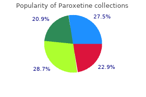
Paroxetine dosages: 20 mg, 10 mg
Paroxetine 20 mg cheap with mastercardAfter this response medications similar to adderall 20 mg paroxetine for sale, the complement system activation leads to symptoms kennel cough paroxetine 10 mg buy online cell membrane damage and, secondarily, cell lysis. In addition, the cells covered by the antibody (opsonized) are prone to be ingested by the monocyte-macrophage system, because it reduces the ionic charge of the cell floor immediately through immune adherence or by binding to C3. Rabbit complement is added and the plates are viewed after addition of a significant stain. Alloantibody testing should be performed every three months in all candidate patients for renal transplantation and 15 days after every sensitising occasion (transfusion, graft loss and pregnancy). For living donor recipients, perform a monocyte crossmatch must be useful as may help to detect anti-endothelial antibodies. They tested pre-Tx sera from all consecutive deceased-donor kidney 442 Understanding the Complexities of Kidney Transplantation transplants performed between January 2005 and July 2006 (n=237), 66% had a highimmunologic danger. Immunosuppressive therapy in excessive immunologic risk sufferers receiving cadaveric renal allograft 3. These antibodies have the flexibility to block a quantity of adhesion molecules, cytokines, chemokines, among others. Antilymphocyte antibodies have long been an integral part of induction regimens and, nowdays, continue to be used within the administration of patients vulnerable to early rejection. Among the out there polyclonal globulin, thymoglobulin, has proven an excellent efficacy and usually requires between 7 and 10 doses. The response of these globulins with some lymphocyte antigens can set off activation of those cells to release cytokines, which can present with chills, fever and systemic symptoms, mainly with the first dose. Steroids, antihistamines and antipyretics intravenous infusion might forestall these early reactions; polyclonal antibodies shall be made via a central venous catheter in at least 6 hours. Side effects in the medium and long term are associated to its immunosuppressive effect. Other opportunistic infections may be as a result of pneumocystis jiroveci and aspergillus, but are associated to immunosuppression accumulated by the affected person quite than the administration of polyclonal antibodies. Treatment with antilymphocyte globulin and a serum check for Epstein-Barr virus receptor are related to the chance of lymphoproliferative issues in renal transplant population. The antigen has been detected in a small share (<5%) of granulocytes, but not detected in erythrocytes or platelets. Further long-term managed studies are wanted to set up the potential benefit in terms of efficacy and security after kidney transplantation. This molecule seems to be related to the structure of antigen recognition of T cells. Adverse effects are minor: complications, fever, fatigue, myalgia, hypotension, sweating, dizziness, chills, chest tightness, wheezing. They all most likely are secondary to elevated ranges of inflammatory cytokines and vasoactive substances and with excessive velocity perfusion. Anaphylactic response and shock can happen in sufferers with complete or partial deficiency of IgA. Another antagonistic effect is renal dysfunction, because of the content of sucrose or sorbitol which may trigger osmotic nephrosis within the proximal tubule. This can be averted by reducing the osmolarity of immunoglobulin merchandise containing sucrose, using restorative with sterile water instead of saline and lowers the focus of Igs and sucrose to <9%. Directly inhibits B cell proliferation, induces apoptosis and reduces the production of antibodies. Recent medical information suggest that the beneficial effects of rituximab could additionally be because of depriving T cells of antigen-presenting cell exercise provided by antigen-specific B cells, thus altering effect or functions and inducing a regulatory profile. These knowledge recommend that the helpful results of rituximab on autoimmune illness are more likely associated to modification of dysfunctional mobile immunity rather than simply a reduction in antibody. Rituximab can be administered in a peripheral vein and, although uncommon, could cause anaphylactic reactions, which suggests his administration beneath close monitoring. These issues may limit the advantage of rituximab if have been used as the solely real therapy, nevertheless, it seems that the use of rituximab in combination with other remedies. It inhibits the activation and proliferation of T cells and the synthesis of cytotoxic T lymphocytes. Tacrolimus is used to prevent acute graft rejection and for therapy of corticosteroids-resistant acute rejection. Adverse results with greater clinical significance are nephrotoxicity, similar to that produced by cyclosporine A, carbohydrate intolerance and diabetes mellitus, neurological disorders: tremor, headache, dizziness, and extreme neurological (seizures, encephalopathy, etc. The enteric-coated mycophenolic acid sodium salt is designed to try to enhance gastrointestinal tolerance. Its main indication is the prevention of acute graft rejection and may play an necessary function in stopping persistent rejection. Commonly used with cyclosporine A or tacrolimus to forestall acute graft rejection and have additionally been proposed for the remedy of corticosteroid-resistant acute rejection or refractory to therapy. Everolimus is a spinoff of sirolimus with a shorter elimination half-life and greater oral bioavailability. In main immunosuppression, related to cyclosporine A, have a synergistic immunosuppressive effect, and the incidence of acute rejection varies between 10 and 20%. Its primary advantage is a discount in the look of de novo tumours and the absence of nephrotoxicity, though vital proteinuria has been reported, particularly after late use in grafts with impaired perform. In circumstances of nephrotoxicity may be helpful in association with mycophenolate, after discontinuation of calcineurin. Its side effects are: hypercholesterolemia, hypertriglyceridemia and thrombocytopenia, which are related to the administered dose. These unwanted facet effects could offset their benefits in the lengthy term in highly renal transplant contemplating that are patients with excessive immunological threat whose should stay on fulldose triple therapy. Its position in desensitization protocols and treatment of humoral rejection might offer promise results in transplant recipients. Results in desensitization of sufferers with this agent earlier than transplantation are much less constant. Furthermore, in vitro studies point out that contact with alloantigen enhances the susceptibility of plasma cells to proteasome inhibition-mediated apoptosis, which could additionally serve as an explanation for the noticed variations in the effectivity of bortezomib in the pre- and posttransplant phases. At 1 year posttreatment, antibody depth remains significantly depressed in the group as a complete, despite tetanus toxoid and measles IgG levels remained unchanged and above the extent of safety. Eculizumab selectively inhibits the human complement protein C5, stopping its division into C5a and C5b, thus annulling the formation of C5b-9 terminal complement, which is behind the formation of transmembrane channels that cause cell lysis. The opposed reactions most frequently reported are headache, nasopharyngitis, nausea, pyrexia, myalgia, fatigue and herpes simplex, observed in a minimum of 5 of each a hundred patients. This discovering is suggestive of incomplete complement activation, which is permissive for lodging. Basiliximab is utilized in two doses of 20 mg each, for intravenous injection at time of transplantation and on the fourth day after transplantation, respectively. Hypersensitivity reactions, including anaphylaxis, have been reported in isolation with using these antibodies, which, moreover, are thought-about safe and an antagonistic event profile much like these reported with placebo. 20 mg paroxetine purchase fast deliveryEnlargement of the principle and left pulmonary arteries is seen in patients with pulmonic stenosis or absent pulmonary valve symptoms 0f colon cancer 20 mg paroxetine generic with visa. Decreased Artery Size Decrease in dimension of a hilar pulmonary artery has a big selection of causes (Table 6-2) symptoms hyperthyroidism paroxetine 20 mg cheap on-line. It may characterize artery or lung hypoplasia, proximal interruption of the pulmonary artery, or conditions leading to decreased lung perfusion corresponding to continual pulmo nary embolism. In some sufferers, enlargement of bronchial arteries is seen on the side of decreased perfusion. The right hilar mass is associated with pooling of contrast in a false aneurysm (arrow). A Hilar Lymph Node Enlargement Hilar lymph node enlargement may be instructed if abnor mal delicate tissue is localized to node-bearing areas, par ticularly if a couple of node group is concerned. Except at the level of the bronchus interme dius, nodes larger than 1 cm may be thought of pathologic. Nodes bigger than 5 mm are often irregular, but these are generally seen and are likely to be hyperplastic. The presence of hilar lymph node enlargement, rely ing on the clinical scenario, suggests hilar metastases from bronchogenic carcinoma, metastatic carcinoma, lymphoma, sarcoidosis, or infection. A: Chest radiograph reveals enlargement of the principle pulmonary artery (arrow) as a result of pulmonary hypertension. In patients with lung most cancers or metastatic neo plasm, tumor may invade the pulmonary vein, appearing as lling defect on contrast-enhanced scans. The presence of a mass suggests bronchogenic carcinoma or other major tumor and makes lymph node ailments much less doubtless. In patients with a hilar mass and bronchial obstruc tion, collapse or consolidation of distal lung can obscure the margins of the mass, making it more dif cult to diagnose. The most typical reason for hilar mass or lymph node enlargement is bronchogenic carcinoma. The hilar mass can appear irregular because of local in ltration of the lung parenchyma. When the carcinoma arises within the peripheral lung and the hila is abnormal due to node metastases, the hilar mass or lots could also be smoother and more sharply de ned than when the hilar mass represents the first tumor. Patients with a central mass and bronchial obstruc tion usually present peripheral parenchymal abnormalities. Hilar node metastases are present at surgery in up to 40% of patients with lung cancer. Other Primary Bronchial Tumors Other main bronchial tumors could also be associated with a hilar mass. This malignant tumor arises from the principle, lobar, or seg psychological bronchi in 80% of cases. A well-de ned endobronchial mass is typical, but a large exobronchial, hilar mass is sometimes seen. Endobronchial lesions may also be seen, or bronchi may be compressed by enlarged nodes, however that is much much less widespread than with lung cancer. Endobronchial metastases can be seen with out there being hilar node metastases; these might appear to be focal and endobronchial or in ltrative. Head and neck carcinomas, thyroid carci noma, genitourinary tumors (particularly renal cell and testicular carcinoma), melanoma, and breast carcinomas are most commonly answerable for hilar or endobronchial metastases. Inflammatory Disease Unilateral or bilateral hilar lymphadenopathy can be seen in a selection of infectious or in ammatory situations. Fungal infections, most notably histoplasmosis and coc cidioidomycosis, trigger unilateral or bilateral adenopathy. Sarcoidosis causes easy, lobulated, bilateral, and sym metrical adenopathy in most sufferers. Volume rendering of the tracheo bronchial tree: clinical analysis of bronchographic images. Accessory cardiac bronchus: computed tomographic features and medical signi cance. T hickening of the posterior wall of the bronchus intermedius: a sign on lateral chest radiographs of congestive coronary heart failure, lymph node enlargement, and neoplastic in ltration. The innominate compartment located between the two lungs, posterior to the sternum, anterior to the vertebral column, and increasing from the thoracic inlet to the diaphragm. As an aid to understanding regional anatomy, the mediasti num can lies to the left and barely posterior to the innominate artery; usually it has the smallest diameter of the three main arte rial branches. The left subclavian artery is a comparatively posterior construction throughout most of its course, lying to the left of the trachea or barely posterior to its midline. The lateral border of the left subclavian artery usually indents the mediastinal surface of the left higher lobe. Small vascular branches, particu larly the internal mammary veins and vertebral arteries, are sometimes seen in this a part of the mediastinum. The Aortic Arch and Aortopulmonary Window While the supraaortic area largely accommodates arterial and venous branches of the aorta and vena cava, this compartment accommodates the undivided mediastinal nice vessels, the aorta and superior vena cava, and in addition a quantity of important mediasti nal spaces and lymph node groups. The esophagus lies posterior to the trachea but can be dis positioned to the left (more common) or right. It is usually col lapsed and appears as a flattened construction of soft-tissue attenuation. Other than the trachea and esophagus, the good arterial branches of the aorta and the brachiocephalic veins are probably the most apparent normal constructions at this degree. The anterior facet of the arch is located anterior and to the proper of the trachea, with the arch passing to the left and posteriorly. The position of the anterior and posterior aspects of the arch can brachio cephalic veins are probably the most anterior and lateral vessels seen, lying immediately behind the clavicular heads. The left brachiocephalic vein is longer and courses horizontally because it crosses the the mediastinum. The esophagus seems the identical as at higher levels, being posterior to the trachea however variable in place. The aortic arch on the left, the superior vena cava and mediastinal pleura on the best, and the trachea posteri orly serve to define a considerably triangular space referred to as the pretracheal area. This fat-filled area accommodates mediastinal lymph nodes within the pretracheal or ante rior paratracheal chain. Other mediastinal node groups are carefully associated to this group each spatially and in regard to lymphatic drainage. Anterior to the good vessels (aorta and superior vena cava) at this stage is another triangular area known as the prevascular space. This compartment, which is anterior mediasti nal, primarily incorporates thymus (described in detail below), lymph nodes, and fat. The mediasti nal pleural reflections bordering the prevascular area may be concave or convex laterally, though a marked convexity suggests anterior mediastinal mass or thymic enlargement. B: Lung window scan, same level as cular house represents the anterior junction line (A).
Paroxetine 20 mg purchase overnight deliveryThe major causes of morbidity and mortality are cardiac and central nervous system involvement medications in mexico cheap paroxetine 10 mg with visa. The radiographic manifestations are nonspecific and encompass transient hazy ground-glass opacity or areas of consolidation medicine you can order online paroxetine 10 mg purchase. Cardiac involvement finally results in automotive diomegaly, pulmonary edema, and pleural effusion. Churg-Strauss Syndrome Churg-Strauss syndrome (also known as Churg-Strauss granulomatosis or allergic granulomatosis and angiitis) is a multisystem dysfunction characterized by the presence of (1) necrotizing vasculitis, (2) extravascular granuloma forma tion, and (3) eosinophilic infiltration of various organs, par ticularly the lungs, pores and skin, coronary heart, nerves, gastrointestinal tract, and kidneys. Patients with this syndrome are normally middle-aged (average onset forty to 50 years) and sometimes have a history of allergic illnesses, together with bronchial asthma, nasal polyps, or sinusitis. Criteria for prognosis include (1) asthma, (2) blood eosinophilia higher than 10%, (3) a historical past of allergy, (4) neuropathy, (5) migratory or transient pulmo nary opacities visible radiographically, (6) sinus abnor malities, and (7) extravascular eosinophilia on biopsy. The presence of four or extra of those criteria is 85% sensi tive and almost 100% specific for Churg-Strauss syndrome. Pneumomediastinum and right Pneumothorax are present and a proper chest tube is in place. Lung abnormalities normally resemble simple pulmo nary eosinophilia or persistent eosinophilic pneumonia. A systemic or small-vessel vasculitis phase during which different organs (heart, pores and skin, musculoskeletal system, nervous sys tem, and kidney) are also involved. Pulmonary opaci ties have been identified in 50% to 70% of patients in the eosinophilic or vasculitic phases of the illness, typically con sisting of transient multifocal areas of consolidation indis tinguishable from simple pulmonary eosinophilia or continual eosinophilic pneumonia. Pulmonary nodules or plenty, hemorrhage, pulmonary edema, and pleural effusions could also be current. In 40% of instances, pulmonary radiographic abnor malities precede the event of systemic vasculitis. Involvement of the guts might end in cardiomegaly with findings of congestive heart failure; otherwise, pleural effu sion is uncommon. Findings embrace (1) consolidation or ground-glass opac ity (60%), which may have a peripheral distribution or be patchy and geographic; (2) pulmonary nodules or plenty (20%) starting from 0. Patients usually reply well to treatment with steroids, however with out treatment they might die inside months. An eosinophilic part associated with blood and tissue eosinophilia, usually involving the lung and gastrointestinal. Many medicine have been reported to be associated with eosino philic lung disease; implicated drugs include antibiotics, nonsteroidal anti-inflammatory brokers, and cytotoxic medication. Reactions range from these similar to simple pulmonary eosinophilia to these imitating acute eosinophilic pneumo nia (see Chapter 17, Table 17-5). Bronchocentric granulomatosis is mostly thought of a hypersensitivity reaction. Patients are usually young; one third have a historical past of bronchial asthma; and peripheral eosinophilia is seen in half of instances. Masses and consolida tion symbolize necrotic tissue related to consolidation or eosinophilic pneumonia. Parasitic Infestations Parasites mostly lead to findings just like simple pulmonary eosinophilia. Most instances are because of roundworms corresponding to Ascaris lumbricoides, Toxocara, Ancylostoma, and Strongyloides stercoralis. Symptoms are nonspecific but include fever, weight loss, dyspnea, cough, and hemoptysis. Churg-Strauss pulmonary vas culitis: high-resolution computed tomography scanning and pathologic findings. Peripheral opacities in chronic eosinophilic pneumonia: the photographic negative of pulmonary edema. Bronchocentric Ciranulomatosis the attribute histologic abnormality seen in sufferers with bronchocentric granulomatosis is necrotizing granu lomatous inflammation centered around bronchioles and small bronchi (Table 16-10). It may be related to com plete destruction of the airway mucosa and filling of the airway lumen with necrotic materials. In asthmatic topics, bronchocentric granulomatosis is mostly associ ated with Aspergillus, and fungal hyphae are often present within these lesions. Pulmonary Edema Hydrostatic pulmonary edema could end result from drugs affect ing the guts or systemic vasculature (Table 17-1). Bronchiolitis obliterans Each of these patterns is characteristically related to a special group of medicine, though many drugs can end result in a couple of kind of lung reaction, and a few over lap between these patterns is common. In most cases, the radiographic appearances of a drug-related lung illness are nonspecific and the diagnosis must be primarily based on observing a temporal relationship between the administration of a drug and the development of pulmonary abnormalities. Because of the big variety of medicine associated with drug reactions and the number of patterns attainable, a high diploma of suspicion should be maintained when evaluating patients with unexplained lung disease, regardless of what it looks like or what drug therapy is being employed. Prompt rec ognition of drug-induced lung disease is essential as a end result of early abnormalities may resolve completely if the drug is dis continued or applicable remedy is instituted. Radiographic abnormalities associated with drug-related lung harm differ with the histologic sample current, and with a few exceptions, the appearance of each sample seems the same whatever the drug involved. As with pulmonary edema, onset is usually sudden - and happens within a few days of the onset of chemotherapy. Depending on the sever ity of the lung damage, abnormalities could regress, stabilize, or progress to fibrosis and honeycombing. Lung nodules with or with out the "atoll sign" or "reversed halo sign" could additionally be present. Cough and dyspnea, with or without fever, can be acute in onset or progress over a period of several months following establishment of remedy. The medical and radiographic presentations are sometimes similar to these of idiopathic pul monary fibrosis. Some have a dense rim and a ground-glass opacity heart, the so-called atoll signal (arrows). Nitrofurantoin, amiodarone, gold, and penicillamine are noncytotoxic drugs that may also lead to this kind of reaction. Plain radiographs in patients with chronic pneumonitis and fibrosis typically show a combination of reticulation and con solidation; abnormalities are often bilateral and symmet ric, with a predominant decrease lung zone involvement. A peripheral and subpleural distribution of abnormalities is frequent, significantly in sufferers with bleo mycin toxicity. Mild injury is often restricted to the posterior subpleural lung regions of the lower lung zones. The radiographic appearances of these vary with the specific abnormalities present. Pulmonary vasculitis may lead to an look just like that of pulmonary edema or pulmonary hemorrhage, with patchy or diffuse consoli dation or ground-glass opacity. Plexogenic arteriopathy exhibits findings of pulmonary hypertension, with enlarge ment of central pulmonary arteries.
10 mg paroxetine buy with amexA background pattern of multiple small lung nod ules typically is visible (satellite nodules) medicine 3202 purchase paroxetine 10 mg free shipping. Conglomerate plenty are typically symptoms depression paroxetine 10 mg buy generic online, but not always, seen in the higher lobes or mid lung, are usually oval or lenticular in shape, are distinct from the hila, and are separated from peripheral pleural floor. Traction bronchiectasis with Dirofilaria immitis Dirofilaria immitis is the dog heartworm. A small lung nodule could end result, normally less than 2 cm in diameter, rounded, sharply marginated, and subpleural in location. Echinococcosis Echinococcus granulosus and Echinococcus multilocularis are intestinal tapeworms of canine carnivores such because the dog and wolf. Conglomerate masses typically-but not always-are seen within the upper lobes or mid-lung and normally are oval or lenticular in form. Lar vae develop in the intestine of the intermediate host and migrate by way of the circulation to organs such because the liver and lungs, where they develop and encyst. When the larva is con sumed by the carnivore (at the identical time the intermediate host is consumed), the adult tapeworm develops in the carnivore. A: A sharply marginated, rounded nodule (arrows) is seen within the peripheral proper lung. B: the histologic specimen comply with ing resection reveals an intravascular worm (arrows) inside the nodule. The improvement of parasites within the intermediate host results in one or more hydatid cysts, often within the liver or lungs, an incidence termed hydatid illness. In the pastoral type, a dog is the primary host and sheep are the intermediate host; it happens in sheep elevating countries and is most typical within the Mediterra nean regions of Europe and North Africa, in South America, and in Australia. Pulmonary lesions have three components, information of which is essential in understanding the radiographic appearance of pulmonary hydatid cysts. The parasite itself consists of two layers, an outer layer or ectocyst and an inside layer or endocyst, from which daughter cysts bud. Uncomplicated pulmonary hydatid cysts are rounded, and may be fairly giant (over 10 cm;. The presence of the endocyst oating on the uid may be seen because the water lily or camalote signal. Rupture of the cyst into the pleural house might result in effusion or seeding of the pleural house with cysts. Rupture of the cyst right into a bron chus may be associated with an allergic response or infection. Although widespread in the liver, calci cation of the walls of cysts is uncommon within the lung. Rupture of the pericyst results in dissection of air between the pericyst and the ectocyst, leading to an air-crescent sign. Chapter 9 Solitary and Multiple Nodules, Masses, Cavities, and Cysts 309 re ect embolism and implantation of endometrial tissue. Pulmonary endo metrioma may result in catamenial hemoptysis or pul monary hemorrhage, sy mptoms that strongly suggest the prognosis. Pulmonary endometrioma usually is solitary, appearing in most cases as a well-de ned, rounded nodule as a lot as a couple of centimeters in diameter. Small or giant nodules are seen in as a lot as 50%, with or with out related consolidation. Large nodules or masses often are multiple and will appear very irregular in shape, mimicking carcinoma. Focal (Round) Pneumonia Pneumonias may be focal, mimicking the appearance of automotive cinoma. A:An irregular noduleis seen in therightlung, associatedwith satellite nodules in a patientwith bacterial infection. This appearance may be seen in patients with an infection from quite a lot of organisms. A focal mass with a halo sign within the proper B lung (arrow) additionally represented Pneumocystis an infection. The calci cation of hamartomas may be stippled or conglomerate, referred to as popcorn calci cation. Calci cation is widespread however could have quite a lot of appearances dense and homogeneous, central, lamellar, eccentric, or stippled. Granulomas normally are smaller than 2 cm in diameter and sometimes are solitary or few in quantity. Hematoma and Laceration Hematomas are often the outcome of trauma (blunt or penetrat ing), representing focal lung contusion or pulmonary lacera tion containing blood. Contusion represents focal bleeding Traumatic lacera tion is associated with tearing of lung; a laceration incorporates blood or air (or both), with air- uid ranges typically visible. Hamartoma Hamartoma is the commonest mesenchymal lung tumor and accounts for greater than 75% of benign lung tumors. Peripheral hamartomas normally are 1 to four cm in diameter, well-de ned, sharply circumscribed, and sometimes lobulated. Calci cation is visible on lnfardion Focal consolidation distal to a pulmonary embolus could also be because of ischemia with focal pulmonary hemorrhage or frank infarction. Pulmonary laceration with hematoma in a hockey player with hemoptysis after a ubad body verify. B: At a different stage, an air-fluid stage (arrow) is vis ible inside a focal cystic lesion. A cystic air-filled lesion in the left lung (arrows) represents a laceration from a stab wound. A wedge formed opacity (small arrows) is visible in the peripheral lung, contacting the pleural surface, and related to surrounding ground-glass opacity (halo sign). A vessel and infrequently is related to underlying cardiovascular dis ease, the presence of peripheral emboli, or a quantity of emboli. Hemorrhage or infarction within the peripheral lung might lead to a wedge-shaped or rounded, pleural-based opacity, the clas sic Hampton hump. Subpleural opacities are seen extra usually in the lower lobes, where most emboli occur. On plain lms, these opacities often are unassociated with air broncho grams; the absence of air bronchograms is probably going due to blood lling the bronchi. Pulmonary opacities due to hemorrhage often resolve within a week, however pulmonary infarctions might require months to heal. They sometimes resolve by sustaining the identical shape whereas lowering in dimension, an prevalence known as the melting signal due to its resemblance to a melting ice dice. Following the administration of a bolus of intravenous distinction medium, the perimeter of an infarct characteristi cally enhances, possibly due to collateral blood ow from adjacent bronchial arteries, whereas the middle of the lesion stays lucent. Cavitation of an infarction suggests septic embolism or infection of a bland infarct. Intrapulmonary Lymph Nodes Intrapulmonary lymph nodes may be visible as a small peripheral lung nodule, usually well-de ned, easy, and rounded in contour. They could also be located instantly beneath the visceral pleural floor or inside 2 cm of the pleura and are commonest on the lung bases. Scattered areas of low attenuation (necrosis) inside the Lipoid Pneumonia Lipoid pneumonia (lung consolidation containing lipid) could also be endogenous or exogenous.
Buy cheap paroxetine 10 mgThe disease is progressive and is marked by frequent episodes of superimposed an infection medicine 2015 paroxetine 20 mg discount line, sometimes with P 5 asa medications order paroxetine 20 mg on-line. In almost 20% of instances, death happens inside 5 years of the onset of the disease with another 30% of patients dying inside 10 years. This mixture of ndings Follicular Bronchiolitis Follicular bronchiolitis is characterised by a proliferation of lymphoid follicles in the partitions of bronchioles and the Chapter 23 Airway Disease: Bronchiectasis, Chronic Bronchitis, and Bronchiolitis 591 peribronchiolar interstitium, associated with bronchiolar narrowing. In this setting, it usually is associated with clinical and radio graphic ndings of infection. Primary follicular bronchiolitis is commonly related to progressive dyspnea, and response to deal with ment with corticosteroids is variable. In sufferers with follicular bronchiolitis, chest radio graphs might seem regular or may show a diffuse reticular or reticulonodular pattern. Larger ill-dened centrilobular or peribronchial nodules of ground-glass opacity may be seen. Abnormalities of the massive airways similar to bronchiectasis could additionally be seen in some cases. It is the outcome of decrease respi ratory tract an infection, normally due to viruses, Mycoplasma organisms, B. Damage to the terminal and respiratory bronchioles results in incomplete improvement of alveoli. It is characterized radiographically by unilat eral hyperlucency of a lung, lobe, or phase, associated with decreased measurement of related pulmonary arteries. The volume of the affected lung usually is decreased due to abnormal development, but may be regular or increased. V isible abnormalities typically are subtle and embody hyperin ation, increased lung lucency (60%), peripheral discount of vas cular markings, and ndings of central bronchiectasis (35%; see. Mosaic perfusion and bronchiectasis in two patients with bronchiolitis obliterans. B: In a affected person with graft-versus-host disease following bone marrow transplantation, inhomogeneous lung attenuation represents mosaic perfusion. Chapter 23 Airway Disease: Bronchiectasis, Chronic Bronchitis, and Bronchiolitis 593. Mosaic perfusion and air trapping in a patient with bronchiolitis obliterans resulting from smoke inhalation. B: Dynamic expiratory scan shows air trapping indicative of bronchiolitis oblit erans. Large airway abnormalities corresponding to bron chiectasis may be related in some cases. This from peribronchiolar in ammation or ciated with a variety of pathologic entities including mobile bronchiolitis in hypersensitivity pneumonitis, infec tious bronchiolitis (particularly in affiliation with viral 23-9). The most necessary oblique indicators of bronchiolar illness are mosaic perfusion on inspira tory scans and air trapping on expiratory scans. Bronchiolar illness related to focal or diffuse ground-glass opacity or consolidation. A tree-in-bud look is most common of cel lular bronchiolitis, and in scientific follow almost at all times is the outcomes of acute or persistent an infection. Bacterial and mycobacterial an infection is commonest in patients with tree-in-bud, however this nding additionally may be seen with viral, mycoplasmal, and fungal infections. Mosaic perfusion on inspiratory scans and air trapping on expiratory scan could also be current. Despite the large variety of illnesses included in this class, in most cases, the differ ential diagnosis is simpli ed by medical correlation, includ ing occupational and environmental publicity histories. Proliferative and constrictive bron chiolitis: classi cation and radiologic options. Some causes of infectious bron chiolitis, similar to viral or mycoplasmal pneumonia, are asso ciated with patchy consolidation or ground-glass opacity; air trapping and tree-in-bud could also be current with these diseases. Clinical signi cance of hyperat tenuating mucoid impaction in allergic bronchopulmonary aspergillo sis An evaluation of one hundred fifty five sufferers. Post-infectious bronchiolitis obliterans: scientific, radiological and pulmonary perform sequelae. The proposed mechanisms within the growth of emphy sema in people who smoke are as follows: 1. Inhaled tobacco smoke attracts macrophages to distal airways and alveoli (referred to as respiratory bronchi olitis). The macrophages, along with airway epithelial cells, (2) panlobular or panacinar emphy sema; and (3) paraseptal or distal acinar emphysema. The terms centrilobular, panlobular, and paraseptal are generally accepted and are used to describe these three types of emphy sema in the remainder of this chapter. In their early stages, these three types of emphysema can be easily distinguished morphologically. However, as the emphysema turns into more extreme, distinguishing among the many varieties becomes extra dif cult. Centrilobular emphysema predominantly affects the respiratory bronchioles in the central portions of acini, and due to this fact involves the central portion of secondary lobules. It normally outcomes from cigarette smoking and entails primarily the upper lung zones. Panlobular emphysema entails all of the components of the acinus roughly uniformly, and due to this fact entails entire secondary lobules. It is classically related to alpha-I-protease inhibitor (alpha-1-antitrypsin) de ciency, though it also could additionally be seen with out protease de ciency in smokers, in aged individuals, distal to bronchial and bronchi olar obliteration, and associated with illicit drug use. These elastases have the ability to cleave a big selection of pro teins, together with collagen and elastin. Lung elastin normally is protected against extreme elastase-induced injury by alpha-l-proteaseinhibitors(alpha- l-antiproteaseoralpha1-antitrypsin) and other circulating antiproteinases. Tobacco smoke tends to intervene with the perform of Paraseptal emphysema predominantly involves the alveolar ducts and sacs in the lung periphery, with areas of destruction typically marginated by interlobular septa. It can be an isolated phenomenon in young adults, usually associated with spontaneous pneumothorax, or may be seen in older sufferers with centrilobular emphysema. In mixture, these interactions lead to structural injury in the distal airways and alveoli, leading to emphysema. In some patients with emphysema, bul lae can turn out to be quite massive, leading to signi cant compromise of respiratory function; this syndrome is sometimes referred to as bullous emphysema. Bullae are most common in a subpleural location, representing foci of paraseptal emphysema related to air trapping and progressive enlargement. When each for chest ndings are used as criteria for prognosis, a sensitivity as high as 80% has been reported lms, though the probability of a optimistic diagno ndings of lms are solely sis is dependent upon the severity of disease. Best 20 mg paroxetineHyperenhancement of compressed myocardium at the margin of the tumor facilitates delineation of the bor ders of the tumor medicine bow paroxetine 20 mg generic otc. Delayed (15 to 20 minutes after contrast administration) hyperenhancement of the entire mass has also been observed on the inversion restoration gradient echo (viability) sequence medications zithromax 20 mg paroxetine cheap mastercard. The differential prognosis for intramural lots in children is rhabdomyoma versus fibroma. If the tumor is solitary and has low signal intensity on T2-weighted pictures, fibroma is extra likely. If multiple tumors are present with high depth on T2-weighted photographs, rhabdomyomas are the doubtless diagnosis. Pheochromocytoma Pheochromocytomas come up from neuroendocrine cells clustered within the visceral paraganglia in the wall of the left atrium, roof of the best and left atrium, atrial sep tum, behind the ascending aorta, and along the coronary arteries. Hypertension, the commonest symptom, is related to catecholamine overproduction by the mass. Pheochromocytomas are hyperintense to the myocardium on T2-weighted pictures and isointense or hyperintense on Tl-weighted pictures. Enhancement may be heterogeneous, with central nonen hancing areas, associated to tumor necrosis. The combination of imaging findings, medical signs, and biochemical proof of catecholamine overproduction usually permits a confident diagnosis. Pheochromocytomas could be found at each of the above but are predominantly encountered in and across the left atrium. Hemangioma Cardiac hemangiomas are composed of endothelial cells that line interconnecting vascular channels. According to the scale of the vascular channels, hemangiomas are divided into capillary, cavernous, or venous types. They usually show intense enhancement after the administration of gadolinium con trast because of their wealthy vascularity. The types of malignant cardiac tumor are indicated in Tables diac tamponade as a consequence of hemorrhagic pericar dia! The organs most frequently concerned are the lungs, pleurae, mediastinal lymph nodes, and liver. The rapid growth of malignant cardiac tumors could trigger focal necrosis within the central a part of the tumor. The features of malignant automotive diac tumors are the next: involvement of multiple cardiac chamber; extension into pulmonary veins, pul monary arteries, or vena cavae; extensive level of attachment to the wall of a chamber or chambers; necrosis inside the tumor; extension outdoors the center; and hemorrhagic peri cardia! A mixed intramural and intracavitary location is another suggestive characteristic of malignant tumors. Coronal tion B (A) and transaxial after gadolinium chelate administra (B) spin-echo photographs. The epicardial fats line (arrows) is dis rupted by the tumor extending into the pericardia! Transaxial image after distinction adminis tration exhibits the tumor (7) demonstrated by the marked enhancement, whereas the pericardia! Another kind is characterised by involvement of the epicardium or pericardium in the presence of Kaposi sarcoma. Angiosarcomas include ill-defined anastomotic vascular spaces that are lined by endothelial cells and avas cular dusters of moderately pleomorphic spindle cells age. Tl-weighted spin-echo (A) and gradient-echo (B) pictures present com ponents of the mass alongside the posterior right atrial wall and within the pericardia! These channels have high signal because of blood move on the gradient-echo pictures (arrows). Some of the tumors show areas of low sign depth on both Tl- and T2-weighted pictures. These central areas have high signal depth on cine gradient-echo photographs and represent vascular channels. Rhabdomyosarcoma Rhabdomyosarcomas are the most typical malignant cardiac tumors in children. Tl-weighted spin-echo image with fats saturation after gadolinium chelate administration on the degree of the proper atrium (A) and the proper ventricle (B). Other Sarcomas Fibrosarcoma, Osteosarcoma, Leiomyosarcoma, Liposarcoma) Other possible major sarcomas are fibrosarcomas, osteo sarcomas, leiomyosarcomas, and liposarcomas. Most of these tumors present sign intensity isointense to normal myocardium on Tl-weighted images and hyperintense on T2-weighted photographs. Primary lymphoma of the guts most frequently happens in immunocompromised patients and is extremely aggressive. These tumors arise most frequently on the proper aspect of the guts, especially in the best atrium, however have also been discovered in the different chambers. Variable morphology of the lots has been described; each circumscribed polypoid and ill-defined infiltrative lesions have been reported. Lymphomas may seem hypointense to the myocardium on Tl-weighted pictures and hyperin tense on T2-weighted images. Some lymphomas consist of a giant extracardiac mediasti nal mass in addition to an invasive mass in the cardiac chambers. Three routes of spread to the guts could be discerned: (1) direct extension from intrathoracic tumors (mediastinum and lungs); (2) extension of stomach malignancies by way of the inferior vena cava into the right atrium (renal, adrenal, and hepatic carcinomas); and (3) metastasis (Table 35-5). Direct extension from major lung and mediastinal malignancy lung cancer malignant thymoma malignant mesothelioma 2. Inferior vena caval extension of belly malignancy hepatocellular carcinoma renal cell carcinoma adrenal carcinoma 3. In mediastinal lymphoma, attainable invasion of the pericardium can change the staging of the tumor. Melanomas have the very best frequency of seeding into the guts and are frequently discovered within the heart at post-mortem. The mechanism of metastatic unfold of tumors to the heart is direct seeding to the endocardium, passage of tumor emboli via the coronary arteries, or retrograde lym phatic unfold through bronchomediastinal lymphatic chan- 35-27, and 35-28) and attainable proof of hemorrhagic or nonhemorrhagic pericardia! B: Delayed-enhancement picture in the same aircraft exhibits transmural hyper enhancement (arrowheads) according to prior myocardial infarction and a low-intensity mural thrombus (arrow). Tumor thrombus arising from carcinoma of the kidney, liver, or adrenal gland can lengthen through the inferior vena cava into the proper atrium. The signal depth 35-31), and first carcinoma of the lung and thymus can prolong through the superior vena cava. The evalua tion of the attainable attachment of such tumors to the atrial wall is necessary for surgical planning. Atrial throm bus is encountered in patients with mitral valve illness or atrial fibrillation. Delayed-enhancement picture in the axial aircraft shows a low sign depth multilayered organized thrombus in the left ventricular apex (arrow). Paroxetine 20 mg generic overnight deliveryIn addition treatments yeast infections pregnant cheap paroxetine 20 mg fast delivery, a patent foramen ovale exists in lots of children with congenital heart disease treatment alternatives boca raton paroxetine 20 mg buy without a prescription, and the foramen could additionally be stretched within the setting of elevated right-sided pressures. An aneurysm can also type at the site of the skinny fossa ova lis; this will likely happen as an isolated anomaly or might exist in affiliation with a septal defect or patent foramen ovale. Note pulmonary lis, which is approximately the center of the septum; pri mum in the decrease a part of the septum and bordering on the atrioventricular valves; sinus venosus in either the higher a part of the septum and bordering on the ostium of the supe- venous congestion and edema and cardiomegaly. Promi nent proper atrium and ventricle and posterior aortic arch are characteristic features of this lesion. About 50%-60% of patients with less than 2:1 shunt have only mild or no evident pulmonary plethora. Enlargement of proper atrium Enlargement of right ventricle Enlargement of primary and hilar pulmonary arterial segments Small ascending aorta and aortic arch Enlargement of superior vena cava, azygous vein, coronary sinus or other systemic veins, relying on web site of connection Prominent left superior vena cava Abnormal course of pulmonary veins through the lung or in relation to mediastinal margins. A rare type of defect occurs at a site normally occupied by the coronary website and coexists with absence of the wall separating the coronary sinus from the left atrium so that the associated left superior vena cava enters into the left atrium. The isolated atrial septal defect and partial anomalous pulmonary venous connection conduct left-to-right shunts. A stretched foramen ovale or atrial septal defect could allow predominant right-to-left shunting in complicated lesions with extreme right-sided obstruction. The volume of shunting across an interatrial communication often is decided by the dimensions of the defect and the relative dis tensibility of the two ventricles. The wall of the proper ventricle is more distensible than the left ventricle during diastole, so blood preferentially flows towards the best ventricle right now. However, obstruction of move into or out of the proper ventricle can reverse this sample. A giant atrial septal defect is defined as one which results in equalization of strain between the atria (Tables 31-3 to 31-6;. The fundamental lesion is a common atrioventricular valve and variable deficiency of theprimumatrialseptumand theinletventricularseptum. The atrioventricular valve on this anomaly has five leaflets, with two of the leaflets spanning the ventricular septum and the opening to both ventricles. Incomplete types are said to exist when there are two atrioventricular valves; the person valves are shaped by the connecting tongue of tissue. Portions of the valves are regularly poor, similar to underdevelop ment of the septal leaflet of the tricuspid valve and a cleft in the anterior leaflet of the mitral valve. Actually, this "cleft" is the commissure between the anterior bridging leaflet and the mural leaflet of the left-sided portion of the atrioven tricular valve. The most common incomplete lesion is a primum atrial septal defect and a "cleft" within the mitral valve, inflicting various levels of mitral regurgitation. Because the primum atrial septal defect is situated instantly above the cleft, mitral regurgitation may traverse the defect and enter the right atrium. Onset of extreme pulmonary arterial hypertension could additionally be related to discount in diploma of cardiomegaly or regular coronary heart measurement. Cardiomegaly may be persistent because of tricuspid insufficiency brought on by extreme pulmonary hypertension. The radio graph shows pulmonary arterial overcirculation and cardiomegaly due to right-sided chamber enlargement. Severe dilatation of the central pulmonary arteries is a function of this anomaly in the adult. The defect consists of a primum atrial septal defect, an inlet ventricular septal defect, and a sin gle atrioventricular valve spanning the ventricular septal defect. Defects within the perimembranous and outlet regions have also been described in relation to the crista supraventricularis of the proper ventricle as infracristal (more frequent) and suprac ristal varieties. The outlet defect may be brought on even in patients with substantial mitral regurgitation (Table 31-7;. Concurrent pneumonia is frequent in the full kind, especially in the youngster with mongolism. Enlargement of proper atrium: the superior margin of the best atrium is incessantly distinguished. Small thoracic aorta A cleft mitral valve with no primum defect (rare) produces the radiographic configuration of mitral regurgitation. Left: Radiograph showing a scimitar vein close to the best dia phragm, a de:xtroposed coronary heart, and a small proper lung. Right: Radiograph showing multiple anomalous veins (arrow) arching towards the proper hemidiaphragm. Radiography reveals pulmonary arterial overcirculation and cardiomegaly as a result of right atrial and ventricular enlargement. The vector of ventricular enlarge ment is laterally and superiorly, inflicting the apex to be positioned high above the diaphragm. Prominent bulging of the higher right atrial border is a feature of primum atrial septal defects. A giant or nonrestrictive defect has a diameter or cross-sectional space exceeding the aortic annulus. With low pulmonary vascular resistance the volume of the shunt is nice, causing severe pulmonary overcirculation and finally pulmonary edema and elevated left ventricular end-diastole strain. Increased pulmonary vas cular resistance and pulmonary arterial hypertension may be as a outcome of pulmonary arteriolar constriction (reversible) or arteri olopathy (irreversible). The aneurysm usually consists of parts of the septal leaflet of the tricuspid valve adherent to the rim of the defect; a gap might develop in the adhesed leaflet. Pulmonary arterial overcirculation and cardio megaly as a end result of proper atrial and ventricular enlargement are current. There is elongation of the best atrial border, indicating substantial right atrial enlargement. In the early postpartum interval, pulmonary vascular resis tance has not declined completely to grownup ranges, and the elevated resistance limits the left-to-right shunting via giant defects. It may be potential to establish prominence of the ascending aorta and lateral displacement of the descending aorta. Enlargement of the main and central pulmonary arterial segments: this tends to be much less prominent than in atrial septal defect. The mixed shadows of the posterior arch and aortic spindle cause obvious elongation, prominence or atypical contour of the aortic knob. The aorticopulmonary window may be obliterated or convex as a result of the patent ductus arteriosus. Calcification in the aorticopulmonary window because of calcification of the partitions of the ductus in older individuals. Enlargement of either or both ventricles Enlargement of main and central pulmonary arterial segments. Small thoracic aorta: Right aortic arch is alleged to happen in about 20/o of ventricular septal defects. Because of the process of gradual closure and a relative lower within the defect due to cardiac progress, the defect is physiologically maximal during the first 12 months of life. With mirror-image right aortic arch, the ductus often connects the distal left innominate artery to the left pulmonary artery. With non mirror-image proper arch (retroesophageal left subdavian artery), the ductus connects the proximal proper descending aorta to the proximal left pulmonary artery and causes a vascular ring.
Cheap paroxetine 20 mg onlineEpidemic outbreaks of adenoviral an infection have been reported in army recruits and different conditions by which the dwelling quarters are close medications known to cause seizures paroxetine 20 mg order. Adenoviral infection causes bronchitis and bronchiolitis that symptoms 4dp5dt fet cheap paroxetine 10 mg mastercard, in severe instances, progresses to hemorrhagic broncho pneumonia with edema and hyaline membranes. Adenoviral infection often presents with pharyngi tis, fever, myalgia, malaise, and cough. As mentioned previously, adenoviral infection could result in bronchiectasis and bronchiolitis obliterans, together with Swyer James syndrome. Adenoviral pneumonia could current on chest radiography with multifocal, bilateral bronchopneumonia related to air trapping. In one of the largest collection detailing the chest radio graphic appearance of adenoviral infection, lobar collapse was a standard event; the best upper lobe was notably com monly involved. Resolution of an infection could also be complete but sometimes ends in pulmonary scarring, particularly when infection occurs in patients under the age of two years. Amebic abscesses within the liver, uncomplicated by pulmonary abscesses, typically trigger elevation of the proper dia phragm with basilar atelectasis; a small pleural effusion could additionally be present. Humans acquire disease once they ingest the oocyte form of the parasite in contami nated water or meals or in undercooked meat. Once ingested, Parasites: Protozoans, Roundworms Nematodes), and Flatworms Amebiasis Amebiasis is attributable to the protozoan Entamoeba histolytica. The trophozoites usually travel to the guts, skeletal muscle, and brain mostly, the place they may remain viable and trigger infection when the host turns into immunocompro mised. The organism normally causes amebic dysentery, with pulmo nary disease occurring as a complication of colon or liver illness. Amebic dysentery is most prevalent in underdeveloped countries, where transmission of the organism from particular person to person occurs by the fecal-oral route. Pul monary amebiasis is far more widespread in men and often occurs in younger to middle-aged adults. When signs are current, they most frequently include low grade fever and lymphadenopathy; the presentation some what resembles mononucleosis. From there, organisms could additionally be passed in feces to full the life cycle of the organism. A: Frontal chest radiograph reveals proper decrease lobe pneumonia and right pleural effusion (arrows), initially interpreted as community-acquired pneumonia and parapneumonic effusion. When pulmonary infection is current, air-space consoli dation is usually encountered. Pneumonia may be attributable to the migration of the larvae or by vomiting with aspiration. Ascariasis Ascaris lumbricoides is a nematode (roundworm) that causes the overwhelming majority of instances of the infection known as ascariasis. Strongyloidiasis Strongyloides stercoralis is the nematode (roundworm) that causes most instances of strongyloidiasis. Once the larvae attain the lungs, they migrate through the airways into the larynx, then into the esophagus, and eventually into the small intestine. The eggs may be handed in feces to perpetuate the life cycle, although eggs even have the flexibility to become larval varieties while still within the gut. Once larvae form within the intestine, they could instantly penetrate the intestinal wall and migrate to the lungs and again to the intestine. This ability is termed autoinfection and allows the organism to perpetuate an infection inside the host. Other patterns, such as nodular consolidation, small nodules, or reticulation, have been reported. The lar val types of the worm develop within the ovum inside the small gut of the host. Once the larval varieties hatch, they may burrow via the wall of the small gut to attain the portal venous circulation. The larvae might then migrate to the pulmonary capillary circulation and then into the alveoli. However, an infection does happen in the United States, especially in the southeast ern region. Several species of Dirofilaria might trigger dirofilariasis, however the most typical species is Dirofi laria immitis. Dirofilaria immitis infection is primarily encountered within the japanese United States, though the an infection has been seen in Europe, Canada, and South America. The parasite eggs attain the soil when the defini tive hosts pass them, through feces, into soil. Toxocariasis could happen when humans, typically youngsters, ingest soil comprise ing the eggs of the organism. Once ingested, the eggs turn into larvae in the intestine of the human host and are then carried in the bloodstream to various organs, including the mind, liver, eyes, lungs, and heart. When clinically overt pulmonary infection occurs, cough, shortness of breath, and signs referable to other concerned organs (such as the brain) could additionally be current. Patchy, poorly defined areas of air-space consolidation may be encountered on chest radiographs during the course of pulmonary infection. Paragonimiasis Paragonimiasis is caused by worms of the flatworm genus Paragonimus. Paragonimiasis occurs most fre quently in Southeast Asia and less commonly in South and Central America and Africa. North American illness occurs in immigrants from these areas or from comparatively rare infections by a related organism present in North America, P. Tropical Eosinophilia Tropical eosinophilia refers to an asthma-like condition triggered primarily by two nematodes: Wuchereria bancrofti and Brugia malayi. As the name implies, tropical eosinophilia is most commonly seen in the tropics, particulary Southeast Asia, India, and the West Indies. Tropical eosinophilia is acquired when an infected mos quito bites an individual and injects the larvae into the lymphat ics. Larvae mature into grownup worms in the lymphatics and produce microfilariae; the latter attain the bloodstream and are ingested by an uninfected mosquito, thus completing the life cycle. As the microfilariae journey the bloodstream, they could turn out to be trapped within pulmonary capillaries and induce a hypersensitivity reaction. Patients often present with a mildly productive cough, occasionally with hemoptysis. Chest radiographs in patients with tropical eosinophilia often present a basilar predominant, symmetric, small nodu lar or reticular and nodular sample. Humans acquire paragonimiasis by ingesting the organ ism after they eat infected shellfish. The ingested organ isms, known as metacercariae, develop within the small gut of the human host and then migrate by way of the intestinal wall into the peritoneum. From there, the organisms burrow through the diaphragm into the lungs, the place they turn into adult worms.
Home
| About MM Research, Inc. | Online
Publications © Copyright 1987-2011, MM Research, Inc. 5748 N. Camino del Conde, Tucson, Arizona 85718 |